|
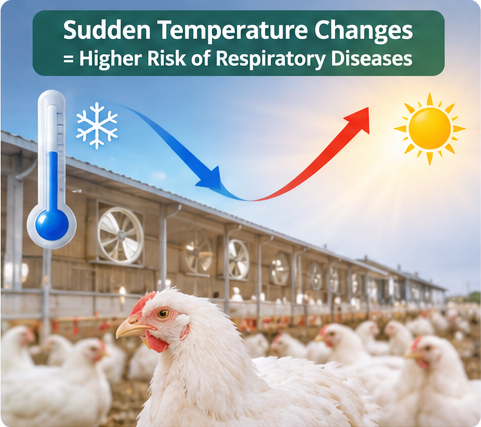
Respiratory Diseases in Poultry: 5 Field Cases & SolutionsWhen daytime and nighttime temperatures fluctuate sharply—whether during autumn-winter transitions in temperate zones, rainy seasons in the tropics, or cool desert nights—respiratory diseases in poultry quickly become the primary driver of flock mortality, medication costs, and lost egg production. For farm managers, veterinarians, and animal health distributors, the biggest challenge is rarely a single pathogen. Instead, cold stress and reduced house ventilation trigger a cascade: high ammonia levels and dry or chilled air paralyze the mucociliary escalator in the chicken trachea. Once that physical barrier fails, primary viruses, Mycoplasma, and opportunistic bacteria like Escherichia coli turn a mild snick into a costly poultry respiratory disease complex (PRDC). Rather than listing textbook definitions, this guide examines five real-world field case scenarios Representing the most common seasonal respiratory threats: Low Pathogenic Avian Influenza (H9N2), Infectious Bronchitis (IB), Infectious Coryza, Infectious Laryngotracheitis (ILT), and Chronic Respiratory Disease (CRD). By walking through the clinical presentation, necropsy findings, and root-cause corrective actions in each case, you can build a more accurate field diagnosis and a sustainable prevention program.
Quick Reference: Differential Diagnosis of 5 Major Poultry Respiratory DiseasesBefore diving into the individual farm investigations, use this field matrix to compare the hallmark signs of the five conditions when examining a flock.
5 Real-World Field Cases: Diagnosis and Farm-Level ResolutionCase 1: The "Silent" H9N2 Trigger Behind a Broiler Bronchial Plug OutbreakThe Farm ScenarioA commercial broiler operation housing 28,000 birds per shed experienced a sharp nighttime temperature drop of 11°C at 24 days of age. To conserve heat, the farm staff closed 60% of the minimum ventilation inlets. Within 72 hours, birds exhibited wet coughing, sneezing, and increased nasal and ocular discharge. Initially, mortality remained low (0.15% per day), leading the farm manager to suspect a simple chill. However, by Day 29, daily mortality spiked past 1.2%, and feed intake dropped by 22%. (Note: In a similar seasonal case involving a 40,000-bird layer farm at 260 days of age, the initial respiratory rales were accompanied by a sudden laying drop from 92% to 71% within ten days, alongside a surge in sandpaper-shelled and soft-shelled eggs.) Necropsy & Diagnostic Investigation
Post-mortem examination of 15 dead and moribund broilers revealed severe diffuse tracheal mucosal congestion. Crucially, yellowish-white, firm caseous plugs were lodged at the syrinx and primary bronchi—the classic cause of acute asphyxiation. Fibrinous pericarditis, perihepatitis, and airsacculitis were present in over 70% of the necropsied birds. RT-PCR swabs confirmed Low Pathogenic Avian Influenza H9N2, complicated by secondary Mycoplasma gallisepticum and Escherichia coli. Why It Happened & How It Was ResolvedStandalone H9N2 rarely causes massive mortality under ideal laboratory conditions, but on commercial farms, it damages respiratory cilia and induces local immunosuppression. Coupled with elevated house ammonia (>25 ppm from restricted ventilation), it opened the door for E. coli and Mycoplasma to form fatal bronchial casts. • Environmental Correction: Minimum ventilation timers were immediately restored to purge ammonia and moisture while supplemental heaters maintained target house temperature. • Medical & Supportive Intervention: Because bronchial obstruction and viral replication were driving mortality, the veterinary team implemented a three-pronged drinking-water protocol: targeted botanical antiviral and antipyretic support to reduce viral shedding and fever, expectorant respiratory relief to help dissolve tracheal mucus and restore airway patency, and a targeted E. coli bacteriophage application to control the secondary colibacillosis without adding kidney stress. Mortality declined by Day 4 of the intervention. Case 2: Early Chick Chilling, Infectious Bronchitis, and the "False Layer" TrapThe Farm ScenarioA layer enterprise noticed that a flock of 18,000 hens at 210 days of age had plateaued at a 73% laying rate—well below the breed standard of 94%. Strangely, the flock looked externally vibrant: combs were bright red and waxy, pubic bones were wide, vents were moist, and daily feed consumption matched peak targets. Yet, a significant percentage of the eggs collected had rough, corrugated ("wrinkled") shells, thin calcifications, and watery albumen. Tracing the Root Cause (The Brooding History)When the consulting veterinarian audited the flock’s brooding records, the culprit emerged at 6 days of age. During the first week of brooding, a heater malfunction during a cold front had caused huddling, gasping, and wet tracheal rales across the chick house. Mortality had reached 3.5% over five days, with some dead chicks showing swollen, pale, urate-filled kidneys. The chicks had recovered clinically after electrolytes and broad-spectrum antibiotics were given, so no viral diagnostics had been run at the time. Necropsy & Diagnostic ConfirmationExamining 20 non-laying hens (identified by cloacal palpation and cage tracking) revealed fully active ovaries with normal hierarchical follicles. However, the oviducts were severely underdeveloped, segmented, or distended into large, thin-walled cysts filled with clear fluid. Paired serology and historical records confirmed early Infectious Bronchitis Virus (IBV) infection during the first two weeks of life—creating permanent oviduct damage known clinically as False Layer Syndrome. Key Takeaway for ProducersAntibiotics have zero effect on IBV. When young chicks (<14 days old) experience cold stress, maternal antibodies alone cannot protect the delicate respiratory and reproductive epithelium against field IBV variants. • Prevention Protocol: Ensure hatchery or Day-1 spray vaccination includes both classical (e.g., Mass-type) and locally prevalent variant IBV strains. Maintain strict brooding temperatures (no cold drafts) during the first 14 days, and support mucosal and systemic immunity during vaccine windows so chicks neutralize the virus before it reaches the developing oviduct. Once a hen becomes a false layer at maturity, the anatomical damage is irreversible; culling non-layers is the only economic recourse. Case 3: Lightning-Fast Facial Swelling in a Multi-Age Layer Complex (Infectious Coryza)The Farm ScenarioFollowing a week of cold, damp winds, a 50,000-bird multi-age layer farm reported an explosive respiratory outbreak in House 3 (38-week-old layers). Within 48 hours of the first birds showing clear nasal discharge and head-shaking, over 40% of the house exhibited dramatic, firm swelling of the face and infraorbital sinuses—often forcing one or both eyes shut. In males kept in a neighboring breeder row, swollen wattles were prominent. Feed intake plunged by 35%, water consumption dropped, and egg production fell from 91% to 64% inside a single week, though overall mortality remained under 1.5%. Differential Diagnosis & Lab FindingsIn the field, facial swelling can be confused with Swollen Head Syndrome (avian metapneumovirus + E. coli), chronic fowl cholera, or severe mycoplasmosis. However, the extreme speed of spread (24 to 72 hours across an entire house) and a characteristic foul, sticky nasal exudate pointed strongly toward Infectious Coryza. Sinus swabs collected from acutely affected birds (before medication) confirmed Avibacterium paragallinarum. Farm-Level ResolutionRecovered birds remain lifelong carriers of A. paragallinarum, which is why cold-season stress repeatedly triggers outbreaks on multi-age farms when older carrier flocks infect newly housed pullets. • Biosecurity & Vaccination: The farm segregated staff and equipment between age groups and revised its pullet vaccination program to include a trivalent (Serovars A, B, and C) inactivated Coryza vaccine administered twice prior to transfer. • Flock Recovery: Alongside veterinarian-prescribed antimicrobial therapy based on sensitivity testing, drinking water was acidified with a buffered organic acid blend (formic, acetic, and propionic acids) to sanitize the water lines—preventing rapid horizontal transmission of A. paragallinarum through shared nipple drinkers—while botanical respiratory and immune enhancers were used to accelerate feed intake and oviduct recovery. Case 4: Acute Gasping and Bloody Mucus on Feathers (Infectious Laryngotracheitis)The Farm ScenarioIn late autumn, a farm raising 15,000 replacement pullets (16 weeks old) and 30,000 adult layers experienced sudden deaths in birds that appeared to be in prime body condition. Upon entering the house at dawn, the farm manager heard loud, high-pitched wheezing and moist rales. Affected birds sat on their hocks and stretched their necks forward and upward with every inhalation ("pump-handle" or "star-gazing" respiration), gasping violently through open beaks. Blood-stained mucus was splattered on cage wires, feed troughs, and the wing feathers of neighboring birds. Daily mortality escalated rapidly. Necropsy & Differential AlertOpening the larynx and trachea immediately revealed the cause of suffocation: severe hemorrhagic tracheitis with free blood clots, mucoid bloody casts, and yellowish diphtheritic membranes completely occluding the upper airway. (Veterinary Caution: Diphtheritic/wet fowl pox can also form yellowish plugs in the larynx that look nearly identical to subacute ILT on necropsy; PCR or histopathology looking for intranuclear inclusion bodies confirms Infectious Laryngotracheitis Virus [ILTV].) Emergency Containment & Airway ManagementBecause birds with ILT die primarily from mechanical asphyxiation rather than systemic organ failure, aggressive handling or stressful spraying can accelerate death. • Immediate Containment: Movement between houses was locked down. Under veterinary supervision, emergency vaccination was performed in unaffected houses to establish competitive protection before field virus spread. • Soothing the Trachea: In the affected house, keeping the birds calm, improving air quality, and administering a concentrated botanical bronchodilator and anti-inflammatory oral solution in drinking water helped soothe tracheal hemorrhage, reduce laryngeal edema, and loosen fibrinous plugs so birds could expel them without suffocating. Case 5: The Chronic Profit Drain—Mycoplasma gallisepticum (CRD) Turning Into Severe ColibacillosisThe Farm ScenarioA 20,000-bird broiler breeder rearer flock in a region with warm days (28°C) and chilly nights (13°C) developed a persistent, low-grade respiratory rattle. Every morning, workers noticed birds shaking their heads, flicking nasal mucus, and displaying tiny, soap-like foam bubbles in the medial corner of the eyes. Mortality barely moved (0.04% daily), so the issue was ignored for two weeks. Following a routine grading and handling stress at 10 weeks, mortality quadrupled, uniformity collapsed, and birds showed severe lethargy and greenish-white diarrhea. Necropsy FindingsEarly cases showed only mild catarrhal sinusitis and cloudy, beaded air sacs. However, birds dying after the handling stress displayed thick, "cheesy" yellow exudate filling the thoracic and abdominal air sacs, along with classic fibrinous perihepatitis and pericarditis ("frosted liver and heart"). Serology and bacterial culture confirmed Chronic Respiratory Disease (CRD) driven by Mycoplasma gallisepticum (MG), complicated by secondary avian pathogenic Escherichia coli (APEC). Breaking the CRD–E. coli CycleStandalone Mycoplasma is a slow-moving pathogen that thrives whenever cold drafts hit the birds. Yet its real economic devastation comes from stripping the respiratory epithelium and inviting E. coli into the bloodstream and air sacs. • Fixing the Micro-Environment: Smoke-pencil testing revealed cold air dropping directly onto the birds' backs during nighttime minimum ventilation cycles because inlet static pressure was too low. Adjusting baffle openings directed cold incoming air along the roof peak, allowing it to warm before reaching bird level. • Targeted Intervention: To avoid driving multidrug resistance through repeated antibiotic courses, the farm combined targeted anti-mycoplasmal management with a drinking-water E. coli bacteriophage cocktail to lyse pathogenic E. coli specifically, alongside herbal respiratory care to clear air-sac exudates. A Practical 4-Step Protocol for Cold-Season Respiratory Control
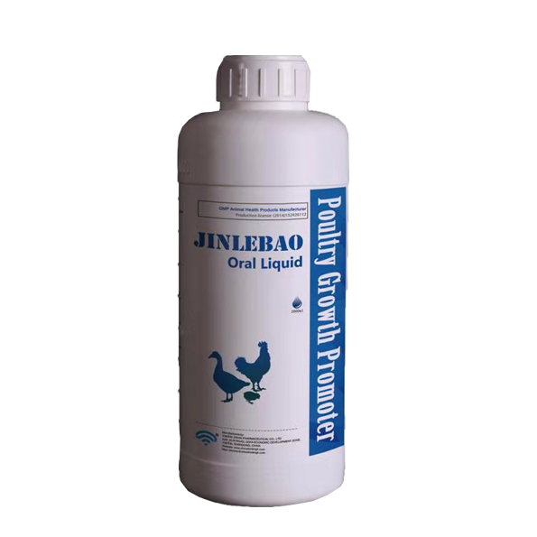
Whether you manage farms in Southeast Asia, the Middle East, Africa, or Latin America, seasonal respiratory outbreaks follow predictable rules. Use this four-step framework to protect flock performance: 1. Balance Heat Retention with Minimum VentilationNever seal a poultry house completely to save heating fuel. When ammonia exceeds 15–20 ppm or relative humidity climbs above 70%, tracheal cilia lose their ability to sweep out dust and viruses. Use negative-pressure inlets properly so cold outside air mixes with warm air near the ceiling before dropping to litter level. 2. Audit Vaccine Timing—and Protect the Immune WindowLive respiratory vaccines (ND, IB, ILT) work by replicating mildly in the respiratory mucosa. If a live vaccine is administered while the flock is chilled, incubating Mycoplasma, or immunosuppressed, the vaccine reaction itself can trigger a full-blown respiratory complex. Support innate and adaptive immunity around vaccination windows. 3. Stop Pathogen Transmission in the Drinking LineDuring respiratory outbreaks, birds dip their beaks—coated with nasal mucus containing H9N2, IBV, Avibacterium, or Mycoplasma—into drinkers or nipple cups. Unsanitized water lines with biofilm quickly become a super-highway for horizontal infection. Maintaining drinking water pH around 4.0 during risk periods helps suppress bacterial proliferation in the water system. (Always pause acidifiers and sanitizers before and during live oral vaccinations according to label instructions.) 4. Use Multi-Target, Residue-Free Supportive ToolsAs global poultry markets restrict heavy antibiotic reliance, combining precise biosecurity with botanical medicines, bacteriophages, and organic acids allows veterinarians to address viral pressure, airway inflammation, and secondary bacterial infections simultaneously without withdrawal-period penalties. Where Targeted Botanical Support Fits in a Respiratory ProgramIn field investigations like the cases above, respiratory losses usually stem from two simultaneous pressures: primary viral replication (such as H9N2 or IBV) and physical airway obstruction (mucus, bronchial plugs, and tracheal inflammation). Alongside ventilation management, biosecurity, and veterinary diagnostics, Yantai Jinhai Pharmaceutical Co., Ltd. provides two complementary, GMP-manufactured botanical oral solutions designed specifically for seasonal respiratory management:
Frequently Asked Questions (FAQ)Why do poultry respiratory diseases spike when day and night temperatures fluctuate?Sharp temperature drops trigger cold stress, which causes vasoconstriction in the respiratory mucosa and elevates stress hormones (corticosterone). At the same time, farmers often reduce ventilation to keep houses warm, causing ammonia, dust, and humidity to rise. Together, these factors paralyze the tracheal cilia and allow resident or incoming viruses and bacteria to invade the lungs and air sacs. Can hens recover normal egg production after Infectious Bronchitis (IB)?If adult laying hens contract IB, egg production and shell quality typically drop for 3 to 6 weeks and then partially recover, though often remaining 5%–10% below pre-infection peaks. However, if female chicks are infected with certain IBV strains during the first 1 to 2 weeks of life, the virus permanently destroys the developing oviduct, creating "false layers" that ovulate normally at maturity but can never lay normal eggs. How can I tell the difference between Infectious Coryza and CRD in the field?Look at the speed of onset and facial lesions. Infectious Coryza (Avibacterium paragallinarum) spreads explosively across a house within 24–72 hours and causes dramatic, firm swelling of the face and infraorbital sinuses with foul-smelling nasal discharge. CRD (Mycoplasma gallisepticum) develops slowly over weeks, characterized by persistent tracheal rales ("gurgling"), head shaking, and tiny foamy bubbles in the corner of the eye, unless complicated by secondary E. coli. |